Cranial nerves
Introduction
Section titled “Introduction”There are a total of 12 cranial nerves, two of which originates from the cerebrum while the rest originates in the brainstem. The cranial nerves are numbered by their location on the brainstem (superior o inferior, then medial to lateral) and the order of their exit from the cranium (anterior to posterior).
| Cranial nerve | Name | Origin | Function Type |
|---|---|---|---|
| CN I | Olfactory nerve | Cerebrum | Sensory |
| CN II | Optic nerve | Cerebrum | Sensory |
| CN III | Oculomotor nerve | Midbrain-pontine junction | Motor |
| CN IV | Trochlear nerve | Midbrain | Motor |
| CN V | Trigeminal nerve: - Opthalmic - Maxillary - Mandibular | Pons | Both |
| CN VI | Abducens nerve | Pontine-medulla junction | Motor |
| CN VII | Facial nerve | Pontine-medulla junction | Both |
| CN VIII | Vestibulocochlear nerve | Pontine-medulla junction | Sensory |
| CN IX | Glossopharyngeal nerve | Medulla | Both |
| CN X | Vagus nerve | Medulla | Both |
| CN XI | Accessory nerve | Medulla | Motor |
| CN XII | Hypoglossal nerve | Medulla | Motor |
Cranial Nerves and Medical Imaging
Section titled “Cranial Nerves and Medical Imaging”Each cranial nerve is described below briefly and shown in different image modalities.
Olfactory nerve
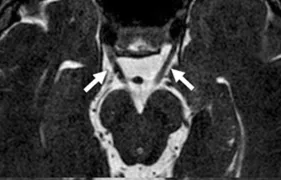
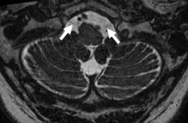
Section titled “Olfactory nerve”Consists of white matter tracts not surrounded by Schwann cells. Enters the anterior cranial fossa through the cribriform plates and terminate in the olfactory bulb. The nerve courses between the gyrus rectus and medial orbital gyrus. The secondary axons terminate in the inferomedial temporal lobe, uncus, and entorhinal cortex.
A Steady-state free precession (SSFP) axial image showing the olfactory nerves (arrows).
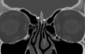
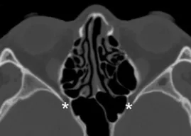
Coronal plane image of cribriform plate (asterisks).
Optic nerve
Section titled “Optic nerve”Emerges from the posterior pole of ocular globe and divided into four segments:
- intraocular
- intraorbital
- intracanalicular
- prechiasmatic
The optic nerve from each side passes through its respective optic canal and joins to form the optic chiasm. Then optic tract courses along the cerebral peduncles and synapses at the lateral geniculate nuclei after which optic radiation reaches the primary visual cortex in the occiptal lobe (Brodmann area 17).
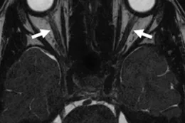
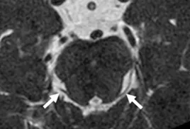
A Steady-state free precession (SSFP) axial image showing the optic nerves (arrows).
Optic canal (asterisks) and superior orbital fissure (arrow)
Also see: Annotated MRI of Optic pathway from Radioepaedia
Oculomotor nerve
Section titled “Oculomotor nerve”Somatic and Parasympathetic functions:
- Somatic motor fibers (innervates inferior superior, middle rectus, inferior oblique, and levator palpebrae superior muscles) originate from nuclear complex located in midbrain at level of superior colliculi, ventral to the cerebral aqueduct and the periaqueductal gray matter.
- Parasympathetic fibers arise in Edinger-Westphal nucleus situated dorsally to the oculomotor nuclear complex.
The nerve emerges from interpeduncular cistern running through the perimensencephalic cistern superiorly to the posterior cerebral artery (PCA) and inferiorly to the superior cerebellar artery (SCA). It then enters the cavernous isnus where iti s the most superior nerve to reach the orbit through superior orbital fissure. Passes through superior orbital fissure.
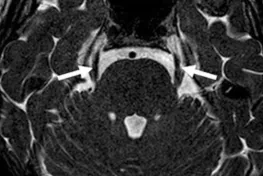
A Steady-state free precession (SSFP) axial image showing the oculomotor nerves (arrows).
Trochlear nerve
Section titled “Trochlear nerve”Originates from the posterior part of midbrain, then runs anteriorly and inferiorly within the subarachnoid space before piercing the dura mater adjacent to the posterior clinoid process of the sphenoid bone. Then it moves along the lateral wall of cavernous sinus before entering the orbit of eye via superior orbital fissure to innervate the superior oblique muscle.
A Steady-state free precession (SSFP) axial image showing the trochlear nerves (arrows).
Trigeminal nerve
Section titled “Trigeminal nerve”The nerve emerges straight forward from the lateral pons and pass in the Meckel’s cave where the trigeminal ganglion is located, then splits into three subvisions:
- Opthalmic nerve (CN V1) Passes through lateral wall of cavernous sinus and exits the skull via superior orbital fissure.
- Maxillary nerve (CN V2) Passes through lateral wall of cavernous sinus and exits the skull via foramen rotundum and crosses the pterygopalatine fossa and then enters the orbits via the inferior orbital fissure, pases within the infraorbital groove, and reaches the face through infraorbital foramen.
- Mandibular nerve (CN V3) exits the skull via formane ovale and passes in the infratemporal fossa where it divides into other nerves.
Terminology
Section titled “Terminology”- Meckel’s cave: a cerebrospinal fluid-containing dural pouch in the middle cranial fossa that contains the trigeminal nerve between prepontine cistern and cavernous sinus and situated in posterolateral aspect of cavernous sinus on either side on sphenoid
A Steady-state free precession (SSFP) axial image showing the trigeminal nerves (arrows).
Axial CT of foramen rotundum (asterisks).
Also see:
- Annotated coronal MRI of trigeminal nerve. Radioepaedia case contributed by Frank Gaillard
Abducens nerve
Section titled “Abducens nerve”Originates at the pontomedullary junction, crosses prepontine cistern in a dorsal to ventral direction, descends near posterior of clivus, passes Dorello canal and enter cavernous sinus, then passes superior orbital fissure to innervate lateral rectus muscle.
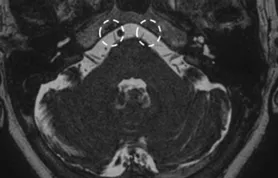
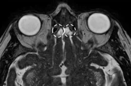
A Steady-state free precession (SSFP) axial image showing the abducens nerves (circles).
Facial nerve
Section titled “Facial nerve”Consists of two portions:
- Proper VII nerve
- Intermediate nerve
Motor nucleus is located at the lower part of pons and motor fibers exit from the lateral portion of pontomedullary sulcus. It then traverses the cerebelopontine cistern and enters the temporal bone through internal acoustic meatus. After passing through the facial canal of the petrous bone, it spits off into three segments:
- labyrinthe
- tympanic
- mastoid
The nerve exits the skull base at stylomastoid foramen and reaches the parotid gland. The mastoid portion seperates into nerve of stapedius and chorda tympani. Afferent sensory fibers of first 2/3 of tongue via chorda tympani to geniculate ganglion Parasympathetic fibers:
Termonology
Section titled “Termonology”- Geniculate ganglion
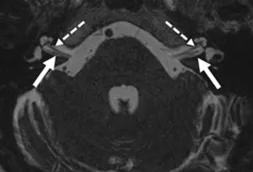
A Steady-state free precession (SSFP) axial image showing the facial nerves (dotted arrows).
Vestibulocochlear nerve
Section titled “Vestibulocochlear nerve”Passes thorugh internal acoustic meastus.
See Facial nerve for MRI image.
Glossopharyngeal nerve
Section titled “Glossopharyngeal nerve”Mixed sensory, motor and secretory nerve. Emerges from the lateral medulla into lateral cerebellomedullary cistern where it is closely associated with flocculus of cerebellum.
Exits the skull via jugular foramen -> enters carotid space -> turns lateral to carotid artery and the stylopharyngeal muscle and continues lateral to the Palatine tonsil area
Branches:
- Lingual at posterior sublingual space to innervate posterior third of tongue (taste and sensory)
- Pharyngeal (sensation from posterior oropharynx and soft palate)
- Sinus nerve (parasympathetic supply to carotid body and sinus)
- Stylopharyngeus (Stylopharyngeus muscle)
- Tympanic branch (Jacobson nerve), sensory information from. Middle ear
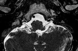
A Steady-state free precession (SSFP) axial image showing the glossopharyngeal and vagus nerves (circles).
Vagus nerve
Section titled “Vagus nerve”Emerges from the lateral medulla, enters the lateral cerebellomedullary cistern and then exits the skull via jugular foramen. The nerve then forms the superior ganglion and inferior vagal ganglion. It descends vertically in the retrostyloid space together with carotid artery to join the upper mediastinum where it gives off the recurrent laryngeal nerve. The nerve enters the abdominal cavity through the esophageal hiatus.
MRI: See Glossopharyngeal nerve.
Accessory nerve
Section titled “Accessory nerve”Two components, spinal and cranial:
- Spinal: Forms from C1-C5/6 nerve roots then ascends towards the foramen magnum, exits the skull through jugular foramen, after which the spinal part then innervates sternonucleidomastoid muscles and trapezius.
- Cranial: Originates from medulla oblongata and shortly after exiting the skull, combines with the vagus nerve at inferior ganglion. Considered part of the vagus nerve.
A Steady-state free precession (SSFP) axial image showing the spinal accessory nerves (circles).
Hypoglossal nerve
Section titled “Hypoglossal nerve”Emerges from medulla oblongata as series of rootlets extending from ventrolateral sulcus of the medulla into the lateral cerebellomedullary cistern near to vertebral artery and PICA, then exits the skull through canal. runs medial to the glossopharyngeal, vagus, and accessory nerves, deeply to the digastric muscle, to innervate a large part of extrinsic and intrinsic tongue muscles
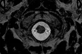
A Steady-state free precession (SSFP) axial image showing the hypoglossal nerves (arrows).
Passageways of the Skull
Section titled “Passageways of the Skull”Optic Canal & Superior orbital fissure
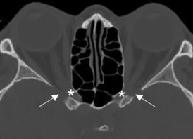
Section titled “Optic Canal & Superior orbital fissure” Axial CT of superior orbital fissure (arrows) and optic canal (asterisks).
Foramen Rotundum
Section titled “Foramen Rotundum” Axial CT of foramen rotundum (asterisks).
Foramen Ovale
Section titled “Foramen Ovale”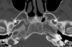
Axial CT of foramen ovale (arrows).
Facial canal
Section titled “Facial canal”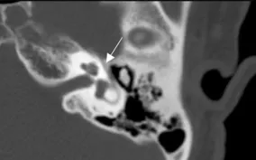
Axial CT of facial canal (arrows).
Internal auditory meatus
Section titled “Internal auditory meatus”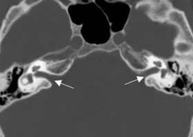
Axial CT of internal auditory meatus (arrows).
Jugular foramen
Section titled “Jugular foramen”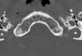
Axial CT of jugular foramen (asterisks).
Hypoglossal canal
Section titled “Hypoglossal canal”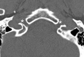
Axial CT of hypoglossal canal (asterisks).
Sources
Section titled “Sources”- Romano, Nicola, Margherita Federici, and Antonio Castaldi. “Imaging of Cranial Nerves: A Pictorial Overview.” Insights into Imaging 10, no. 1 (2019): 33. https://doi.org/10.1186/s13244-019-0719-5.
- Malhotra, Ajay, Long Tu, Vivek B. Kalra, et al. “Neuroimaging of Meckel’s Cave in Normal and Disease Conditions.” Insights into Imaging 9, no. 4 (2018): 499–510. https://doi.org/10.1007/s13244-018-0604-7.